Specialized Ophthalmology in Seoul · Dedicated International Patient Concierge
MOHW Registered · Foreign Patient Care
en-US

Retina & Glaucoma Treatment · Needs Clinic Confirmation
A.I. Diagnostic System
An AI Diagnostic System uses artificial-intelligence algorithms to analyze ophthalmic images or diagnostic measurements and identify patterns that may be associated with eye diseases.
Schedule a ConsultationA.I. Diagnostic System
Medically reviewed by Dr. Kim Yoon Kyung, Director, Ophthalmologist (Master of Medicine), board-certified, Korean Ophthalmological Society
An AI Diagnostic System uses artificial-intelligence algorithms to analyze ophthalmic images or diagnostic measurements and identify patterns that may be associated with eye diseases. At Jamsil Samsung Eye Clinic in Songpa-gu, Seoul, AI has been studied for detecting conditions such as diabetic retinopathy, glaucoma, and age-related macular degeneration from retinal images and other clinical data, and is intended to support clinicians rather than independently replace an ophthalmologist's diagnosis.
What Is the A.I. Diagnostic System?
This system adds a computational layer to conventional ophthalmic diagnosis rather than replacing it.
High-resolution digital fundus cameras, optical coherence tomography (OCT) scanners, cloud-based deep learning algorithms, and automated pattern-recognition software integrated with electronic health record platforms make up the underlying technology.
Autonomous AI algorithms process high-definition retinal or corneal scans in real time, comparing structural vessel patterns, optic nerve parameters, and macular thickness against validated databases to generate automated risk scores or triage flags for clinical review.
Patients seek AI-assisted diagnostic evaluations for rapid, non-invasive screening of retinal conditions like diabetic retinopathy, earlier detection of eye diseases before noticeable symptoms appear, and accelerated review times during routine eye exams.
AI output should not be considered a definitive diagnosis without appropriate clinical assessment; the system is generally intended to support clinicians rather than independently replace an ophthalmologist's diagnosis.
Potential applications include screening or assessment for retinal disease, diabetic retinopathy, glaucoma-related changes, and other image-detectable abnormalities, with final suitability depending on the clinician and the specific AI system used.
Who Oversees AI-Assisted Diagnostics at Jamsil Samsung Eye Clinic

Dr. Kim Yoon Kyung, a director at Jamsil Samsung Eye Clinic, is a board-certified ophthalmologist who graduated from Soonchunhyang University College of Medicine and earned a Master's degree in ophthalmology from the Graduate School of Soonchunhyang University, completing her ophthalmology specialist training at Soonchunhyang University Hospital. Her academic publications include work on open-angle glaucoma and trans-lamina cribrosa pressure, flash electroretinography and visual evoked potentials in diabetic retinopathy with vitreous hemorrhage, and spectral-domain OCT findings in Vogt-Koyanagi-Harada disease — directly relevant to interpreting the retinal and glaucoma-related imaging this AI system analyzes. She is a full member of the Korean Ophthalmological Society, Korean Optometric Association, Korean Contact Lens Society, Korean External Ocular Diseases Society (KEEDS), and Korean Society of Cataract and Refractive Surgery.
Who Might Benefit from AI-Assisted Diagnostics
Patients undergoing ophthalmic screening or diagnostic evaluation where digital imaging or measurable clinical data can be analyzed by an AI-assisted system.
Patients being screened or assessed for retinal disease, diabetic retinopathy, glaucoma-related changes, or other image-detectable abnormalities.
There is no universal exclusion category, since this is a diagnostic-support technology rather than a treatment; it may be less useful as a standalone approach when image or data quality is inadequate, or when a suspected condition falls outside the system's validated indications.
AI output is not intended as a definitive diagnosis on its own without a clinician's assessment, so no patient should expect the AI analysis alone to replace an ophthalmologist's examination.
What to Expect: The Process and Results Timeline
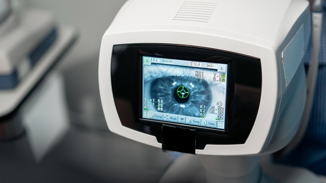
The process follows six steps: the doctor reviews the patient's symptoms and medical history; eye images or other diagnostic measurements are obtained; the AI system analyzes the collected data; the system may flag patterns or findings requiring attention; the ophthalmologist reviews the AI output together with the examination results; and the doctor explains the findings and recommends monitoring or treatment if necessary. No anesthesia is normally required for the AI analysis itself, since it is a software-based analysis of diagnostic data; if the underlying examination involves imaging such as retinal photography or OCT, these are generally non-invasive procedures in their own right.
AI analysis can generally produce results during or shortly after the diagnostic examination, depending on the system, though the clinical interpretation and final diagnosis should come from the ophthalmologist rather than the algorithm. AI analysis does not itself provide a permanent result, and patients may require repeat examinations over time because eye conditions can develop or progress. The AI component may add little time to an existing diagnostic examination, but total appointment time depends on the eye examinations and imaging required; follow-up visits depend on the patient's diagnosis and risk level rather than the AI analysis itself.
Preparation is usually minimal: patients should provide their medical and eye history and inform the doctor about existing eye conditions, medications, and relevant systemic diseases. If pupil dilation is planned as part of the underlying examination, patients may wish to bring sunglasses and arrange transportation in case their vision becomes temporarily blurred. Aftercare for the AI analysis itself generally requires no special instructions beyond those associated with the underlying eye examination; if dilation was performed, patients should wait until vision returns to normal before driving or performing visually demanding activities, and should follow whatever specific aftercare applies to any treatment recommended based on the findings.
Safety, Precision, and Clinic Standards
The AI analysis itself generally causes no physical side effects, since it analyzes diagnostic data rather than performing any procedure on the eye. However, diagnostic errors are possible, including false-positive and false-negative results; poor image quality, differences between patient populations, or conditions outside the algorithm's validated use can affect accuracy, which is why clinical confirmation by the ophthalmologist remains central to every diagnosis made using this technology. The system's automated risk scores and triage flags are intended to support, not substitute for, the same clinical judgment that has always guided ophthalmic diagnosis.
International Patient Considerations
International patients often value the AI Diagnostic System specifically because it can be incorporated into a single, relatively short eye examination visit rather than requiring a separate appointment or a return trip for screening results. Because the underlying imaging — fundus photography, OCT scanning, and related diagnostic measurements — is often already part of a comprehensive eye exam, the AI analysis layer typically does not meaningfully extend the length of a visit already planned for other reasons, which can be useful for patients traveling to Seoul on a limited schedule. Patients should still expect that any finding flagged by the AI system, whether related to diabetic retinopathy, glaucoma-related changes, or macular degeneration, will require the ophthalmologist's own clinical review and, in some cases, additional testing before a treatment plan can be finalized; this clinical review step should be built into travel planning rather than assumed to happen instantly. Patients who travel specifically for a comprehensive screening visit should also ask in advance whether copies of their imaging and AI-assisted analysis can be provided in a format usable by their home-country ophthalmologist, since continuity of retinal and glaucoma monitoring over time is often more clinically valuable than any single screening visit on its own.
Why AI-Assisted Screening Is Being Adopted in Ophthalmology
Retinal and optic nerve disease often progress gradually and without obvious early symptoms, which is part of why conditions such as diabetic retinopathy, glaucoma, and age-related macular degeneration are frequently diagnosed only after some vision loss has already occurred. AI-based pattern recognition has been studied specifically because it can process large volumes of retinal imaging consistently and flag subtle structural changes — such as early microaneurysms in diabetic retinopathy, or small changes in optic nerve fiber layer thickness associated with glaucoma — that may be easy to overlook during a purely visual review, particularly across a busy clinical caseload. This does not mean the AI system detects disease that a trained ophthalmologist would otherwise miss in every case; rather, it is intended to function as a consistency and triage layer, helping direct clinical attention toward images or measurements that warrant closer manual review. The clinical value of this approach depends heavily on the quality of the underlying imaging equipment and the validation behind the specific algorithm used, which is why the system is paired with high-resolution fundus cameras and OCT scanners rather than used as a standalone smartphone-based screening tool.
How This Fits Into a Routine Eye Exam
| Step | What Happens |
|---|---|
| Standard eye exam alone | Ophthalmologist examination, imaging as clinically indicated |
| Eye exam plus AI Diagnostic System | Same exam, plus automated pattern analysis of the imaging obtained, reviewed alongside clinical findings |
Alternatives or complementary approaches to AI analysis include standard ophthalmologist examination, retinal photography, OCT, visual-field testing, intraocular-pressure measurement, and other imaging or diagnostic tests depending on the suspected condition — AI analysis is generally positioned as an adjunct to these established methods rather than a replacement for any of them.
Combining AI Diagnostics with Other Care
Screening with the AI Diagnostic System can be bundled with a general eye exam, allowing retinal and glaucoma-related screening to be added to a routine visit rather than requiring a separate appointment specifically for AI analysis.
The Bottom Line
The AI Diagnostic System analyzes retinal images and diagnostic measurements to flag patterns that may be associated with eye disease, supporting — not replacing — the ophthalmologist's own examination.
It can help with rapid, non-invasive screening for conditions such as diabetic retinopathy, glaucoma-related changes, and macular degeneration.
AI output requires clinical confirmation and is not itself a definitive diagnosis; false-positive and false-negative results are possible.
Dr. Kim Yoon Kyung's academic research spans glaucoma, diabetic retinopathy, and retinal imaging findings, directly relevant to interpreting this system's output.
The AI component adds little time to an existing exam and generally requires no special preparation or aftercare beyond what the underlying eye examination requires.
Frequently Asked Questions
Does the AI system diagnose me, or does my doctor still do that? Your doctor still makes the diagnosis. The AI system analyzes diagnostic images and data to flag patterns that may need attention, but the ophthalmologist reviews this output together with the clinical examination before determining a final diagnosis or treatment recommendation.
Can the AI system make mistakes? Yes — like any diagnostic tool, it can produce false-positive or false-negative results, particularly if image quality is poor or a patient's presentation falls outside the system's validated use, which is why clinical confirmation by your ophthalmologist remains part of every evaluation.
What conditions can this system help screen for? Potential applications include screening or assessment for retinal disease, diabetic retinopathy, glaucoma-related changes, and other conditions that can be detected through imaging analysis, though final suitability depends on your specific case and the clinician's assessment.
Does using the AI system add extra time or cost to my eye exam? The AI component itself generally adds little time to an existing diagnostic examination, since it analyzes imaging that is often already being obtained as part of a comprehensive eye exam; total appointment time depends mainly on the underlying examinations and imaging required.
Do I need to prepare anything special for AI-assisted diagnostics? Preparation is usually minimal — provide your medical and eye history and inform the doctor of existing conditions and medications; if pupil dilation is part of your underlying exam, bring sunglasses and arrange transportation in case of temporary blurred vision.
Book a Consultation at Jamsil Samsung Eye Clinic
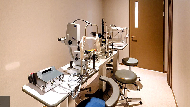
To arrange an eye examination that includes AI-assisted diagnostic screening, contact Jamsil Samsung Eye Clinic in Songpa-gu, Seoul, through its online consultation and contact page or by phone at +82-2-424-7523.
Disclaimer
This page is for general informational purposes and does not constitute individualized medical advice. Diagnosis and treatment recommendations can only be determined through evaluation by a qualified physician; AI-assisted analysis does not replace clinical judgment.
Sources & Further Reading
Other Services
Explore additional ophthalmology treatments offered by our specialists.
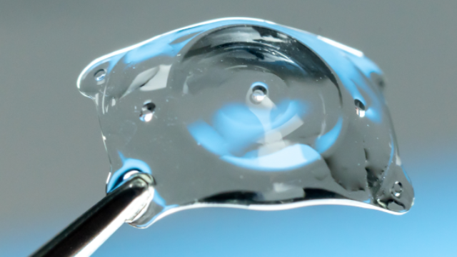
EVO AQUA ICL (Implantable Collamer Lens)
EVO AQUA ICL is a vision-correction procedure in which a specialized corrective lens made of biocompatible Collamer material is inserted directly between the natural lens and the iris, without damaging or reshaping the cornea.
Learn More

Presbyopia Surgery (Clareon PanOptix Pro)
Presbyopia surgery with the Clareon PanOptix Pro trifocal lens corrects presbyopia — the age-related loss of near-focusing ability caused by the eye's lens stiffening and losing elasticity, combined with reduced ciliary-muscle function — by implanting a
Learn More
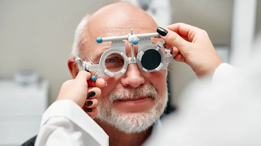
Presbyopia Precision Exam Program
The Presbyopia Precision Exam Program is a specialized diagnostic evaluation designed to thoroughly measure eye health, corneal state, and refractive index before prescribing presbyopia correction or laser vision procedures.
Learn More