Specialized Ophthalmology in Seoul · Dedicated International Patient Concierge
MOHW Registered · Foreign Patient Care
en-CA

Specialty Overview
Retina & Glaucoma Treatment
Retina and glaucoma care at Jamsil Samsung Eye Clinic in Seoul: AI-assisted diagnostics, diabetic retinopathy, macular degeneration, and glaucoma treatment.
Schedule a ConsultationRetina & Glaucoma Treatment
Medically reviewed by Dr. Kim Yoon Kyung, Director, Ophthalmologist (Master of Medicine), board-certified, Korean Ophthalmological Society
Retina and glaucoma care covers comprehensive evaluation, diagnosis, monitoring, and treatment for diseases affecting the retina, vitreous, and optic nerve, including diabetic retinopathy, macular degeneration, glaucoma, and vitreous floaters. At Jamsil Samsung Eye Clinic in Songpa-gu, Seoul, treatment is individualised according to each patient's diagnosis and disease severity, supported by an AI-assisted diagnostic system alongside conventional retinal and glaucoma testing.
What Is Retina & Glaucoma Treatment?
Retina and glaucoma treatment addresses conditions affecting the back of the eye — the light-sensing retina, the vitreous gel that fills the eye, and the optic nerve that carries visual signals to the brain.
Retinal conditions treated include diabetic retinopathy, macular degeneration, retinal tears or detachment, retinal vein occlusion, macular oedema, and vitreous floaters (myodesopsia).
Glaucoma treatment focuses on lowering elevated eye pressure to slow or prevent further optic nerve damage and vision loss.
An AI Diagnostic System supports — but does not replace — the ophthalmologist's clinical judgement by analysing retinal images and diagnostic measurements for patterns associated with disease.
Treatment options range from observation and medication to laser therapy, intravitreal injection, or surgery such as vitrectomy, selective laser trabeculoplasty, or minimally invasive glaucoma surgery (MIGS).
South Korea is recognised internationally for advanced ophthalmic technology and comprehensive diagnostic testing, which is part of why international patients pursue retina and glaucoma evaluation here.
Who Performs Retina & Glaucoma Treatment at Jamsil Samsung Eye Clinic

Dr. Kim Yoon Kyung, Master's degree
Board-certified Ophthalmologist
Dr. Kim Yoon Kyung, a director at Jamsil Samsung Eye Clinic, is a board-certified ophthalmologist who graduated from Soonchunhyang University College of Medicine and earned a Master's degree in ophthalmology from the Graduate School of Soonchunhyang University, completing her ophthalmology specialist training at Soonchunhyang University Hospital. While her primary listed specialisations are orthokeratology (Dream Lens) and vision-correction surgery, her academic research portfolio is directly relevant to retinal and glaucoma disease: her seven supplied publications include work on open-angle glaucoma and trans-lamina cribrosa pressure, flash electroretinography and visual evoked potentials in diabetic retinopathy with vitreous haemorrhage, nestin expression during pharmacologically induced retinal degeneration, anti-VEGF effects on human retinal pigment epithelial cells under high-glucose conditions, and spectral-domain OCT findings in Vogt-Koyanagi-Harada disease — a condition that itself involves inflammatory retinal and choroidal changes. She previously served as Director of Woori Yonsei Eye Clinic and is a full member of the Korean Ophthalmological Society, Korean Optometric Association, Korean Contact Lens Society, Korean External Ocular Diseases Society (KEEDS), and Korean Society of Cataract and Refractive Surgery.
Retina & Glaucoma Services Available
| Service | What It Addresses |
|---|---|
| AI Diagnostic System | AI-assisted analysis of retinal images and diagnostic data to flag patterns associated with disease, reviewed by the ophthalmologist |
| Glaucoma | Diagnosis and pressure-lowering treatment to slow or prevent vision loss from optic nerve damage |
| Diabetic Retinopathy | Detection and management of diabetes-related retinal damage, including macular oedema |
| Macular Degeneration | Evaluation and treatment of central-vision loss from age-related retinal changes |
| Floaters (Myodesopsia) | Evaluation and, when appropriate, treatment of visually significant vitreous floaters |
The AI Diagnostic System uses high-resolution digital fundus cameras, optical coherence tomography (OCT) scanners, and cloud-based deep learning algorithms to compare structural vessel patterns, optic nerve parameters, and macular thickness against validated databases, generating automated risk scores or triage flags that the ophthalmologist reviews together with the clinical examination. Glaucoma treatment uses diagnostic imaging, visual field analysers, and laser systems, with options ranging from medication to selective laser trabeculoplasty or minimally invasive micro-stent implants that enhance natural fluid outflow. Diabetic retinopathy management uses OCT to assess macular swelling and fundus fluorescein angiography to identify leaking vessels, with treatment selected from focal or scatter laser photocoagulation, intravitreal injections, or vitrectomy depending on severity. Macular degeneration care can include precise intravitreal anti-VEGF injections or focal photodynamic laser therapy to seal leaking micro-vessels in the macula. Floaters significant enough to interfere with daily activities can be treated with Nd:YAG laser vitreolysis or, in selected cases, pars plana vitrectomy.
Who Is a Good Candidate for Retina & Glaucoma Treatment
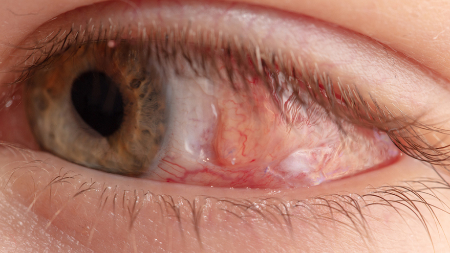
Patients with diabetic retinopathy, retinal tears, retinal detachment, macular degeneration, retinal vein occlusion, or macular oedema.
Patients with glaucoma, elevated intraocular pressure, optic nerve abnormalities, or a family history of glaucoma.
Patients with persistent, visually significant floaters that interfere with reading, computer work, or driving, following a comprehensive retinal examination.
Suitability depends on the specific disease — some patients require only observation or medication rather than laser treatment or surgery, and active systemic illness or uncontrolled medical conditions may delay elective procedures.
Patients whose sudden increase in floaters, new flashes of light, or a curtain-like shadow across their vision should seek urgent evaluation rather than elective treatment, since these can signal a retinal tear or detachment.
Patients without a confirmed diagnosis in one of these categories, or those whose visual symptoms turn out to be caused by a different eye condition entirely, are generally redirected to the more appropriate service — for example, a patient presenting with what appears to be a floater complaint but who is found on examination to have early diabetic retinopathy would be guided toward diabetic retinopathy management rather than floater-specific treatment.
What to Expect: Procedure, Recovery, and Results Timeline
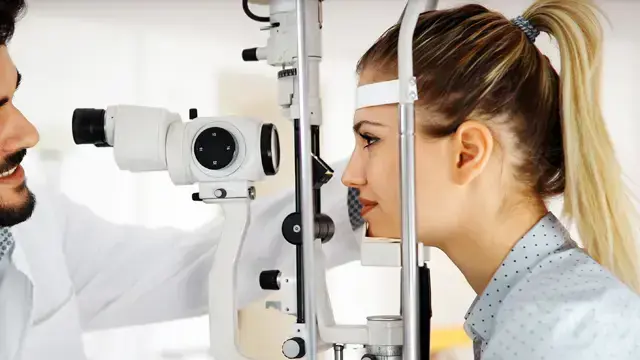
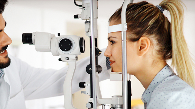
Evaluation begins with a comprehensive eye examination that generally includes visual acuity testing, slit-lamp examination, dilated retinal examination, intraocular pressure measurement, optic nerve evaluation, retinal imaging, and OCT, plus any additional testing indicated by the findings; a first visit with this level of diagnostic testing typically takes about one to two hours. Depending on the diagnosis, the ophthalmologist may recommend observation, medication, laser treatment, an intravitreal injection, or surgery. Most retinal lasers and intravitreal injections are performed under topical anaesthetic eye drops and take about ten to thirty minutes, while vitrectomy or glaucoma surgery may require local anaesthesia with sedation or general anaesthesia and generally takes one to three hours depending on complexity.
Recovery varies significantly by treatment type. Diagnostic evaluation and the AI-assisted imaging itself require little to no recovery, though temporary blurred vision or light sensitivity from pupil-dilating drops can occur until the drops wear off. Laser treatments generally allow a return to normal activities within one to two days. Surgical recovery, such as after vitrectomy or glaucoma surgery, may take several weeks, with vision improving gradually and follow-up visits scheduled to monitor healing. Diagnostic and laser/injection results are typically apparent quickly, but glaucoma treatment in particular is aimed at controlling disease progression rather than restoring vision already lost, and requires lifelong monitoring regardless of which treatment is chosen.
Before any procedure, patients undergo medical history review, medication review, an eye examination, and retinal imaging; blood tests may be required before surgery, and patients should arrange transportation in advance since pupil dilation or surgery can temporarily affect their ability to drive safely. Aftercare across these services generally includes using prescribed eye drops as directed, avoiding rubbing the eye, attending scheduled follow-up visits, monitoring for pain or sudden vision changes, and following any activity restrictions specific to the procedure performed.
Typical visit lengths vary by service and give a useful sense of what to plan for:
| Service | Typical Duration |
|---|---|
| Initial diagnostic consultation | About 1–2 hours, due to comprehensive testing |
| Laser procedures (e.g., SLT, retinal laser) | About 15–30 minutes |
| Intravitreal injection | About 10–15 minutes |
| Vitrectomy or glaucoma surgery | About 1–3 hours, plus post-operative follow-up visits |
Beyond the primary treatment selected, several alternative or complementary approaches exist for most of these conditions, and the treating ophthalmologist selects among them based on diagnosis and severity: for retinal disease, alternatives can include observation, anti-VEGF injections, or vitrectomy; for glaucoma, alternatives can include medication alone, selective laser trabeculoplasty (SLT), minimally invasive glaucoma surgery (MIGS), trabeculectomy, or glaucoma drainage devices, chosen according to how advanced the disease is and how well eye pressure responds to less invasive options first.
International Patient Considerations
International patients pursuing retina and glaucoma care in Korea are often weighing how much diagnostic testing they will need on a single visit, how many procedures or follow-up visits their specific diagnosis will require, and how ongoing monitoring will be handled once they return home. Because conditions such as glaucoma and diabetic retinopathy generally require lifelong or long-term monitoring rather than a single corrective treatment, patients planning treatment abroad should discuss with the clinic, before travelling, how follow-up imaging and pressure checks might be coordinated with an eye care provider in their home country between visits to Seoul. For diagnostic-only visits using the AI Diagnostic System and standard retinal imaging, a single one-to-two-hour appointment is often sufficient to establish a baseline, while active treatment such as vitrectomy or glaucoma surgery should be planned with additional time in Korea for the procedure itself and at least an initial post-operative follow-up visit.
Safety, Precision, and Clinic Standards
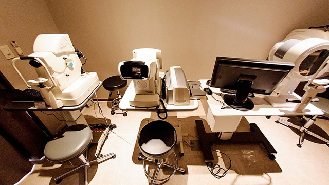
The clinic's retina and glaucoma care is built around advanced retinal imaging and OCT, combined with an AI-assisted diagnostic layer intended to support — not replace — the ophthalmologist's own examination and final diagnosis. Recognised risks vary by treatment: laser and injection-based treatments can cause temporary blurred vision, eye discomfort, or inflammation; vitrectomy carries risks including infection, bleeding, retinal tear or detachment, changes in eye pressure, and cataract progression; and AI-assisted analysis itself carries a risk of false-positive or false-negative findings, particularly where image quality is poor or a patient's presentation falls outside the algorithm's validated use, which is why clinical confirmation by the ophthalmologist remains central to every diagnosis. For floaters specifically, patients are advised to seek urgent care rather than elective treatment if they notice a sudden increase in floaters, new flashes, severe pain, or a curtain-like shadow, since these can indicate a retinal tear or detachment requiring immediate attention.
Because AI-assisted analysis is intended to support rather than replace clinical judgement, the clinic treats any AI-flagged finding as a prompt for further ophthalmologist review rather than as a stand-alone diagnosis, and patients should expect their final diagnosis and treatment plan to come from the treating physician's own examination, not from an algorithm alone.
Combining Retina & Glaucoma Treatment with Other Care
Cataract surgery can be combined with glaucoma surgery in appropriately selected patients.
Vitrectomy can be combined with membrane peeling or retinal laser treatment, or with cataract extraction when clinically indicated.
Floater treatment can be combined with concurrent cataract surgery, peripheral retinal laser photocoagulation to secure retinal tears, or anti-VEGF injections when retinal disease is present at the same time.
The AI Diagnostic System can be bundled into a general eye examination, allowing retinal and glaucoma screening to be added to a routine visit without a separate appointment.
The Bottom Line
Retina and glaucoma care spans diagnosis and treatment for diabetic retinopathy, macular degeneration, glaucoma, and vitreous floaters, individualised to each patient's disease and severity.
An AI Diagnostic System supports faster, more consistent screening but does not replace the ophthalmologist's clinical examination and final diagnosis.
Glaucoma treatment can slow or halt further vision loss but does not restore vision already lost, which is why lifelong monitoring is generally required.
Dr. Kim Yoon Kyung's academic research spans glaucoma, diabetic retinopathy, retinal degeneration, and anti-VEGF therapy, directly relevant to this specialty area.
Sudden new floaters, flashes, or a curtain-like shadow in the vision warrant urgent evaluation rather than a routine appointment.
Frequently Asked Questions
How is glaucoma diagnosed and treated at the clinic?
Diagnosis involves a detailed eye examination, eye-pressure measurement, optic nerve assessment, and visual field testing; treatment is then selected from medication, laser therapy such as selective laser trabeculoplasty, or minimally invasive glaucoma surgery, with the goal of lowering eye pressure to slow disease progression rather than restore vision already lost.
What does the AI Diagnostic System actually do?
It analyses retinal images and other diagnostic measurements using deep learning algorithms to flag patterns that may be associated with disease, such as diabetic retinopathy or glaucoma-related changes, but the ophthalmologist reviews the AI output together with the clinical examination before making any diagnosis or treatment recommendation.
How is diabetic retinopathy treated?
Treatment depends on severity and can include observation, focal or scatter laser photocoagulation, intravitreal injections, or vitrectomy for advanced cases, with OCT and fundus fluorescein angiography used to assess macular swelling and identify leaking blood vessels.
Can floaters be treated, or do I just have to live with them?
Mild floaters that do not significantly affect vision are often simply monitored, but persistent, visually significant floaters can be treated with Nd:YAG laser vitreolysis or, in selected cases, pars plana vitrectomy, following a comprehensive retinal examination to confirm there is no underlying retinal tear or detachment.
What are the warning signs that need urgent attention rather than a routine appointment?
A sudden increase in floaters, new flashes of light, severe eye pain, or a curtain-like shadow across part of the vision should be reported for urgent evaluation, since these can indicate a retinal tear or detachment rather than a condition that can safely wait for a scheduled visit.
Will glaucoma or macular degeneration treatment restore my vision?
Generally, no — glaucoma treatment is aimed at slowing or preventing further vision loss by controlling eye pressure, and macular degeneration treatment is aimed at preserving remaining vision and slowing progression; neither treatment is expected to restore vision already lost, which is why early diagnosis and consistent follow-up matter.
Book a Consultation at Jamsil Samsung Eye Clinic
To arrange a retina or glaucoma evaluation, contact Jamsil Samsung Eye Clinic in Songpa-gu, Seoul, through its online consultation and contact page or by phone at +82-2-424-7523. Patients noticing sudden flashes, a marked increase in floaters, or a curtain-like shadow across their vision should seek urgent evaluation rather than waiting for a routine appointment.
Disclaimer
This page is for general informational purposes and does not constitute individualised medical advice. Diagnosis, treatment selection, and expected outcomes can only be determined through evaluation by a qualified physician, and results are not guaranteed.
Related Services
Ophthalmology procedures available across our clinical departments.
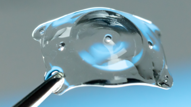
EVO AQUA ICL (Implantable Collamer Lens)
EVO AQUA ICL is a vision-correction procedure in which a specialized corrective lens made of biocompatible Collamer material is inserted directly between the natural lens and the iris, without damaging or reshaping the cornea.
Learn More

Presbyopia Surgery (Clareon PanOptix Pro)
Presbyopia surgery with the Clareon PanOptix Pro trifocal lens corrects presbyopia — the age-related loss of near-focusing ability caused by the eye's lens stiffening and losing elasticity, combined with reduced ciliary muscle function — by implanting a
Learn More
Presbyopia Precision Examination Program
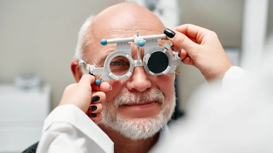
The Presbyopia Precision Examination Program is a specialized diagnostic evaluation designed to thoroughly assess eye health, corneal condition, and refractive index before prescribing presbyopia correction or laser vision procedures.
Learn More