Specialized Ophthalmology in Seoul · Dedicated International Patient Concierge
MOHW Registered · Foreign Patient Care
en-CA


Retina & Glaucoma Treatment · Needs Clinic Confirmation
Macular Degeneration
Age-related macular degeneration (AMD) is a progressive eye condition that affects the macula, the central part of the retina responsible for sharp, detailed vision. It is a leading cause of vision loss among older adults.
Schedule a ConsultationMacular Degeneration

Medically reviewed by Dr. Kim Yoon Kyung, Director, Ophthalmologist (Master of Medicine), certified by the Korean Ophthalmological Society
Age-related macular degeneration (AMD) is a progressive eye condition that damages the macula, the central part of the retina responsible for sharp, detailed vision, and it is a leading cause of vision loss among older adults. At Jamsil Samsung Eye Clinic in Songpa-gu, Seoul, AMD evaluation and management is aimed at detecting the condition early and preserving as much central vision as possible, since damage already present typically cannot be fully reversed.
What Is Macular Degeneration?
Because AMD specifically affects central vision rather than peripheral vision, its symptoms and impact on daily activities differ noticeably from other retinal conditions.
Macular degeneration develops when the macula deteriorates, most commonly as part of the ageing process, and comes in two main forms with different characteristics and treatment approaches.
Dry AMD, the more common form, involves gradual thinning of the macula and the accumulation of small yellow deposits called drusen; it typically progresses slowly over years.
Wet AMD, the less common but more aggressive form, involves abnormal blood vessel growth beneath the retina that can leak fluid or blood, causing more rapid and severe central vision loss if untreated.
Dry AMD can sometimes progress to wet AMD, which is part of why regular monitoring matters even for patients whose condition currently appears stable.
Risk factors include older age, family history, smoking, cardiovascular disease, and high blood pressure; smoking in particular is one of the most significant modifiable risk factors.
Early symptoms can include mild blurring of central vision, difficulty recognising faces, or needing more light for reading; wet AMD can cause more sudden distortion, such as straight lines appearing wavy, or a dark or empty area in the central visual field.
Who Oversees Macular Degeneration Care at Jamsil Samsung Eye Clinic
Dr. Kim Yoon Kyung, a director at Jamsil Samsung Eye Clinic, is a certified ophthalmologist who graduated from Soonchunhyang University College of Medicine and earned a Master's degree in ophthalmology from the Graduate School of Soonchunhyang University, completing her ophthalmology specialist training at Soonchunhyang University Hospital. Her academic publications include research on the effect of anti-VEGF therapy on retinal pigment epithelial cells — directly relevant to wet AMD treatment mechanisms — alongside work on retinal degeneration more broadly and diabetic retinopathy. She is a full member of the Korean Ophthalmological Society, Korean Optometric Association, Korean Contact Lens Society, Korean External Ocular Diseases Society (KEEDS), and Korean Society of Cataract and Refractive Surgery.
Who Should Be Evaluated for Macular Degeneration
Older adults, particularly those over 50, who notice changes in central vision such as blurring, difficulty reading, or trouble recognising faces.
Patients with a family history of AMD, a history of smoking, cardiovascular disease, or high blood pressure, since these factors increase risk.
Anyone noticing sudden visual distortion, such as straight lines appearing wavy or curved, or a new dark or empty area in central vision, should seek prompt evaluation, since this can indicate wet AMD requiring urgent treatment.
Patients already diagnosed with dry AMD who need regular monitoring to detect early signs of progression to wet AMD, which is treatable with more urgency the earlier it is caught.
What to Expect: Diagnosis, Treatment, and Results Timeline
Diagnosis generally involves a dilated eye exam to directly examine the macula, optical coherence tomography (OCT) to assess retinal layers and detect fluid associated with wet AMD, and sometimes fluorescein angiography to visualise abnormal blood vessels. An Amsler grid test may also be used to help detect visual distortion associated with AMD. Treatment differs significantly by type: dry AMD is generally managed through monitoring and, in some cases, specific vitamin and mineral supplementation shown in research to reduce progression risk in certain patients, while wet AMD is typically treated with anti-VEGF injections into the eye, which can reduce abnormal blood vessel activity and fluid leakage. Anaesthesia requirements vary: diagnostic imaging requires none, and anti-VEGF injections are typically performed with topical or local anaesthesia.
Macular degeneration management is generally an ongoing process rather than a single treatment that resolves the condition, particularly for wet AMD, where anti-VEGF injections are often needed on a repeated schedule to maintain treatment benefit over time. Follow-up intervals depend on AMD type and stage: dry AMD monitoring may occur every six to twelve months depending on severity, while wet AMD treatment often begins with more frequent injections, sometimes monthly, before potentially being spaced out as the condition stabilises. Preparation for a dilated eye exam is minimal, though patients should expect temporary blurred vision and light sensitivity afterward. Aftercare after anti-VEGF injections generally involves monitoring for signs of infection or elevated eye pressure and following the specific post-injection instructions provided by the treating ophthalmologist.
International Patient Considerations
International patients being evaluated for AMD should understand that a definitive determination of dry versus wet AMD, and the corresponding treatment plan, generally requires the imaging obtained during an in-person evaluation, so it is difficult to predict in advance which treatment path — if any — will apply before that exam takes place. Patients already receiving anti-VEGF injections for wet AMD should be aware this is typically an ongoing, repeated treatment rather than a single procedure, and should discuss with the clinic in advance how injection scheduling will be managed if their treatment plan calls for injections at intervals extending beyond a single visit to Seoul, including whether coordination with a local retina specialist at home may be appropriate for continuity of care. Bringing prior OCT scans, fluorescein angiography results, and details of any existing AMD diagnosis or treatment history can help the clinic's ophthalmologists assess the current stage and trajectory of the condition more efficiently at the first visit.
Safety, Precision, and Clinic Standards
Diagnostic tools for AMD evaluation generally include OCT scanners, fluorescein angiography equipment, and fundus imaging, along with Amsler grid testing for monitoring central vision distortion, supported by the clinic's AI-assisted diagnostic evaluation described elsewhere in its retina and glaucoma services. Risks associated with anti-VEGF injections include a small chance of infection, elevated eye pressure, or, rarely, more serious complications; dry AMD monitoring and supplementation carry minimal direct risk, though supplementation should be discussed with a doctor given potential interactions or contraindications for individual patients. Because dry AMD can progress to wet AMD without obvious warning, and because wet AMD treatment is generally more effective the earlier it begins, the clinic emphasises prompt evaluation of any new visual distortion or central vision change rather than waiting to see if symptoms resolve on their own.
Living with Central Vision Changes
Even with appropriate medical treatment, some patients with more advanced AMD experience lasting central vision impairment, since damage already present in the macula generally cannot be fully reversed. In these situations, low-vision rehabilitation resources — including magnification devices, adaptive lighting, and specialised reading aids — can help patients maintain independence in daily activities such as reading, cooking, and recognising faces, even though these tools do not restore lost vision itself. Peripheral vision is typically preserved in AMD, since the condition specifically affects the macula rather than the retina as a whole, which is why many patients retain enough peripheral vision to navigate their surroundings safely even when central, detailed vision has been significantly affected. Discussing realistic expectations about visual outcomes, and when appropriate, referral to low-vision support resources, is generally considered part of comprehensive AMD care alongside the direct medical and injection-based treatment of the condition itself.
Comparing Dry and Wet AMD
| Feature | Dry AMD | Wet AMD |
|---|---|---|
| Frequency | More common | Less common |
| Progression | Generally gradual, over years | Can be rapid |
| Underlying process | Macular thinning, drusen deposits | Abnormal blood vessel growth, leakage |
| Typical treatment | Monitoring, sometimes supplementation | Anti-VEGF injections |
| Urgency of new symptoms | Monitor for changes | Sudden distortion warrants prompt evaluation |
Alternative and complementary measures for AMD generally include smoking cessation, blood pressure management, and, for some dry AMD patients, specific vitamin and mineral supplementation discussed individually with a doctor, none of which replace direct monitoring and treatment of the retina itself.
Combining Macular Degeneration Care with Other Treatments

AMD evaluation is frequently combined with glaucoma screening and diabetic retinopathy assessment during the same comprehensive eye exam, since the OCT imaging, dilated exam, and AI-assisted analysis used to evaluate the macula overlap substantially with the diagnostic workup for these other retinal and optic nerve conditions.
Patients newly diagnosed with dry AMD sometimes assume there is nothing that can be done until the condition becomes more severe, but regular monitoring itself serves an active clinical purpose: catching an early transition to wet AMD gives treatment the best chance of preserving central vision, which is why scheduled follow-up remains part of active dry AMD management rather than a passive waiting period.
The Bottom Line
Macular degeneration damages the macula, the retina's central region responsible for sharp vision, and comes in two forms — dry (more common, gradual) and wet (less common, more aggressive).
Sudden visual distortion, such as wavy lines or a new dark spot in central vision, can indicate wet AMD and warrants prompt evaluation.
Dry AMD is generally managed through monitoring and sometimes supplementation; wet AMD is typically treated with anti-VEGF injections.
Smoking is one of the most significant modifiable risk factors for AMD, alongside age, family history, and cardiovascular disease.
Dr. Kim Yoon Kyung's academic research on anti-VEGF effects on retinal pigment epithelial cells is directly relevant to wet AMD treatment mechanisms.
Frequently Asked Questions
Can macular degeneration be cured?
No — AMD is generally a chronic, progressive condition rather than one that is cured. Treatment for wet AMD, primarily anti-VEGF injections, aims to reduce abnormal blood vessel activity and preserve remaining vision rather than restore vision already lost; dry AMD is managed mainly through monitoring and, in some cases, supplementation.
How do I know if I have dry or wet AMD?
This can only be determined through an eye exam that typically includes OCT imaging and sometimes fluorescein angiography; wet AMD often presents with more sudden symptoms such as visual distortion or a new dark spot, while dry AMD tends to progress more gradually, but a clinical exam is needed to confirm which type is present.
What should I do if straight lines suddenly look wavy?
Seek prompt evaluation. Sudden visual distortion, such as straight lines appearing wavy or curved, can indicate wet AMD, which is generally more treatable the earlier it is caught, making timely evaluation important rather than waiting to see if the symptom resolves.
Do vitamins help with macular degeneration?
Specific vitamin and mineral supplementation has been studied for certain dry AMD patients and shown some benefit in reducing progression risk in research settings, though this should be discussed individually with a doctor rather than started independently, since it is not appropriate or beneficial for every AMD patient.
How often will I need anti-VEGF injections if I have wet AMD?
This varies by individual case, but treatment often begins with more frequent injections, sometimes monthly, before potentially being spaced out over time as the condition responds to treatment and stabilises.
Book a Consultation at Jamsil Samsung Eye Clinic
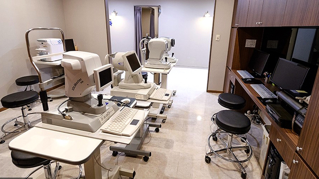
To schedule a macular degeneration evaluation or discuss treatment options, contact Jamsil Samsung Eye Clinic in Songpa-gu, Seoul, through its online consultation and contact page or by phone at +82-2-424-7523.
Disclaimer
This page is for general informational purposes and does not constitute individualised medical advice. Diagnosis and treatment recommendations can only be determined through evaluation by a qualified doctor.
Other Services
Explore additional ophthalmology treatments offered by our specialists.
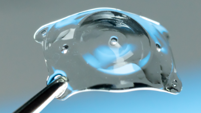
EVO AQUA ICL (Implantable Collamer Lens)
EVO AQUA ICL is a vision-correction procedure in which a specialized corrective lens made of biocompatible Collamer material is inserted directly between the natural lens and the iris, without damaging or reshaping the cornea.
Learn More

Presbyopia Surgery (Clareon PanOptix Pro)
Presbyopia surgery with the Clareon PanOptix Pro trifocal lens corrects presbyopia — the age-related loss of near-focusing ability caused by the eye's lens stiffening and losing elasticity, combined with reduced ciliary muscle function — by implanting a
Learn More
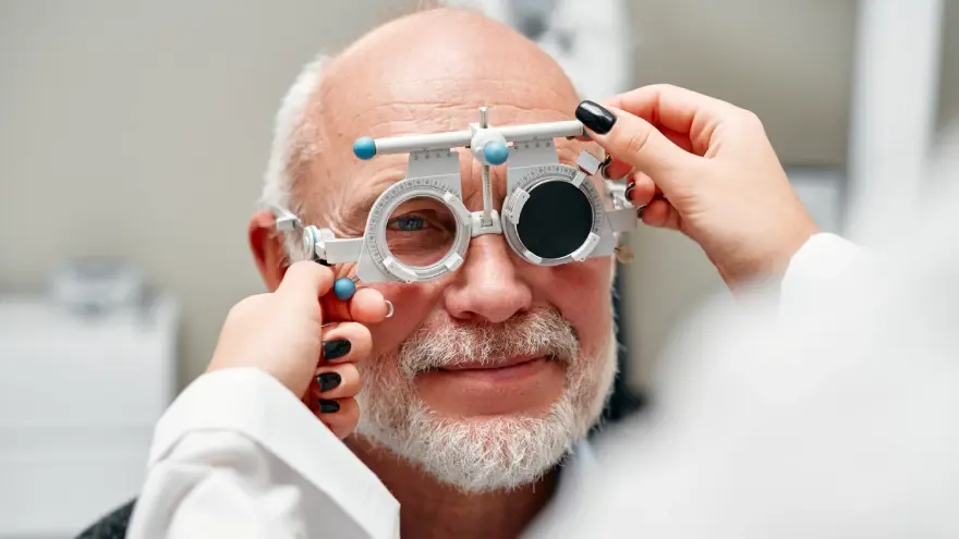
Presbyopia Precision Examination Program
The Presbyopia Precision Examination Program is a specialized diagnostic evaluation designed to thoroughly assess eye health, corneal condition, and refractive index before prescribing presbyopia correction or laser vision procedures.
Learn More