Specialized Ophthalmology in Seoul · Dedicated International Patient Concierge
MOHW Registered · Foreign Patient Care
en-CA


Retina & Glaucoma Treatment · Needs Clinic Confirmation
Floaters (Myodesopsia)
Floaters are shadows cast on the retina by tiny particles suspended in the vitreous gel. They commonly appear as specks, threads, cobwebs, or ring-shaped forms that move with the eye and tend to drift after the eye stops moving.
Schedule a ConsultationFloaters (Myodesopsia)

Medically reviewed by Dr. Kim Yoon Kyung, Director, Ophthalmologist (Master of Medicine), certified by the Korean Ophthalmological Society
Floaters, medically known as myodesopsia, are small specks, threads, or cobweb-like shapes that drift across a person's field of vision, caused by tiny clumps of collagen fibres or cells within the vitreous, the gel-like substance filling the back of the eye. At Jamsil Samsung Eye Clinic in Songpa-gu, Seoul, floaters are most often a benign, age-related change, but evaluation is important because they can occasionally signal a more serious underlying condition, such as retinal tear or detachment, that requires prompt treatment.
What Are Floaters?
Understanding why floaters occur — and why a sudden change in floaters warrants prompt evaluation rather than being dismissed — is central to how the clinic approaches this common complaint.
Floaters are shadows cast on the retina by small particles suspended in the vitreous gel, most commonly appearing as specks, threads, cobwebs, or ring-shaped shapes that move as the eye moves and tend to drift when the eye stops moving.
As people age, the vitreous gel gradually becomes more liquid and can separate from the retina, a process called vitreous detachment, which is a common and usually benign cause of new floaters, particularly after age 50 to 60.
Floaters can also result from inflammation inside the eye, bleeding into the vitreous, or, less commonly, as a symptom accompanying a retinal tear or retinal detachment, which is why any sudden increase in floaters warrants prompt evaluation.
Risk factors for floaters include ageing, nearsightedness (myopia), previous eye surgery such as cataract surgery, eye trauma, and diabetic eye disease.
Most floaters related to normal age-related vitreous changes are harmless and, while they do not disappear entirely, tend to become less noticeable over time as the brain adapts to them.
A sudden shower of new floaters, especially when accompanied by flashes of light or a curtain-like shadow in the peripheral vision, is a recognised warning sign that should prompt urgent evaluation to rule out retinal tear or detachment.
Who Oversees Floaters Evaluation at Jamsil Samsung Eye Clinic

Dr. Kim Yoon Kyung, a director at Jamsil Samsung Eye Clinic, is a certified ophthalmologist who graduated from Soonchunhyang University College of Medicine and earned a Master's degree in ophthalmology from the Graduate School of Soonchunhyang University, completing her ophthalmology specialist training at Soonchunhyang University Hospital. Her academic publications include research on retinal degeneration and diabetic retinopathy with vitreous haemorrhage — both directly relevant to understanding vitreous and retinal causes of floaters — alongside work on glaucoma and macular conditions. She is a full member of the Korean Ophthalmological Society, Korean Optometric Association, Korean Contact Lens Society, Korean External Ocular Diseases Society (KEEDS), and Korean Society of Cataract and Refractive Surgery.
Who Should Be Evaluated for Floaters
Anyone experiencing new floaters for the first time, since an initial dilated eye exam is recommended to confirm the cause and rule out retinal tear or detachment.
Patients with risk factors such as significant nearsightedness, previous eye surgery, eye trauma, or diabetic eye disease, who may have a somewhat higher likelihood of a floater related to a more serious underlying cause.
Anyone noticing a sudden increase in floaters, new flashes of light, or a shadow or curtain affecting part of their vision should seek urgent evaluation, as these can indicate a retinal tear or detachment.
Patients with long-standing, stable floaters who simply want reassurance or an updated evaluation, particularly if it has been some time since their last comprehensive dilated eye exam.
What to Expect: Evaluation, Treatment, and Results Timeline
Evaluation for floaters generally involves a comprehensive dilated eye exam to directly examine the vitreous and retina, looking specifically for retinal tears, holes, or detachment that could explain the symptom; optical coherence tomography (OCT) or ultrasound imaging may be used in some cases for further assessment. Anaesthesia is not required for this evaluation, which is performed with diagnostic drops for dilation. In most cases, once serious retinal causes have been ruled out, floaters from normal age-related vitreous changes do not require treatment and are managed through reassurance and, if needed, periodic monitoring. In rare cases where floaters are dense enough to significantly impair vision and do not improve over time, a vitrectomy — a surgical procedure to remove the vitreous gel along with the floaters — may be considered, though this is generally reserved for more severe, persistent cases given the surgery's own risks.
Most floaters related to normal ageing do not disappear completely but tend to become less bothersome over weeks to months as the brain adapts and the particles settle within the vitreous. If a retinal tear or detachment is identified during evaluation, treatment — which can include laser or surgical repair — is typically time-sensitive and would follow a different, more urgent management timeline than benign floaters. Preparation for the evaluation is minimal, though patients should expect temporary blurred vision and light sensitivity after dilation and should arrange transportation accordingly. No specific aftercare is required for floaters themselves once serious causes are excluded, though patients are generally advised to seek follow-up evaluation if their symptoms change significantly.
International Patient Considerations
International patients presenting with floaters should understand that the primary goal of an initial evaluation is to rule out a retinal tear or detachment, and that this determination generally requires the dilated exam and imaging performed in person, so it cannot be reliably assessed remotely beforehand. Patients whose evaluation confirms benign, age-related floaters typically do not require any further treatment or follow-up beyond routine eye care, which can make this a straightforward, single-visit evaluation for travellers. However, patients who are found to have a retinal tear or early detachment should be prepared for the possibility that treatment may need to begin promptly, which can affect travel plans; discussing this possibility with the clinic in advance, particularly for patients with known risk factors such as high myopia or previous eye trauma, can help set realistic expectations before arrival.
Safety, Precision, and Clinic Standards
Diagnostic tools for floater evaluation generally include dilated fundus examination equipment, OCT scanners, and, when needed, ultrasound imaging of the vitreous and retina, supported by the clinic's AI-assisted diagnostic evaluation described elsewhere in its retina and glaucoma services. The evaluation itself carries negligible risk beyond temporary light sensitivity and blurred vision from dilating drops. If vitrectomy surgery is eventually recommended for severe, persistent floaters, it carries risks similar to other intraocular surgery, including infection, bleeding, retinal detachment, and cataract formation, which is why this option is generally reserved for cases where floaters significantly and persistently impair vision rather than being offered as a routine solution for typical age-related floaters.
Floaters Versus Flashes: Why the Distinction Matters
Patients sometimes use "floaters" and "flashes" interchangeably, but they describe distinct phenomena with different clinical implications. Floaters are visible shapes — specks, threads, cobwebs, or rings — that appear to drift within the field of vision as particles in the vitreous cast shadows on the retina. Flashes, by contrast, are brief streaks or sparks of light, often most noticeable in peripheral vision or in dim lighting, that occur when the vitreous tugs on or separates from the retina. Either symptom alone can occur as part of a benign, age-related vitreous change, but the combination of new floaters together with new flashes is considered a stronger warning sign for retinal tear or detachment than either symptom in isolation, which is why the evaluation process specifically asks about both symptoms together rather than treating them as unrelated complaints.
Comparing Floater Causes and Management
| Cause | Typical Presentation | Management |
|---|---|---|
| Normal age-related vitreous changes | Gradual onset, stable over time | Reassurance, periodic monitoring, generally no treatment needed |
| Vitreous detachment | New floaters, sometimes with flashes | Dilated exam to rule out retinal tear; monitoring if retina intact |
| Retinal tear or detachment | Sudden shower of floaters, flashes, shadow/curtain | Urgent laser or surgical treatment |
| Severe, persistent floaters impairing vision | Dense, unchanging floaters affecting daily activities | Vitrectomy considered in select cases |
Alternative approaches for bothersome but benign floaters generally involve continued monitoring and reassurance rather than active treatment, since most floaters become less noticeable over time without intervention.
Combining Floaters Evaluation with Other Care

An evaluation for floaters is frequently combined with broader retinal screening, glaucoma assessment, and diabetic retinopathy evaluation during the same comprehensive dilated eye exam, since the same examination techniques used to rule out retinal tear or detachment also allow assessment of these other conditions.
Patients are sometimes surprised that a symptom as simple as a few new floaters warrants a full dilated eye exam, but this reflects the clinic's approach that any new floater deserves a baseline evaluation to distinguish between the common, benign cause and the less common but more urgent possibility of a retinal problem — rather than assuming every case is harmless without confirming it first.
The Bottom Line
Floaters are shadows cast by particles in the vitreous gel and are most often a benign, age-related change, particularly after age 50 to 60.
A sudden increase in floaters, new flashes of light, or a shadow or curtain in vision warrants urgent evaluation to rule out retinal tear or detachment.
Most benign floaters do not require treatment and become less noticeable over time as the brain adapts.
Vitrectomy surgery is a treatment option reserved for select cases of severe, persistent floaters that significantly impair vision.
Dr. Kim Yoon Kyung's research on retinal degeneration and vitreous haemorrhage is directly relevant to evaluating vitreous and retinal causes of floaters.
Frequently Asked Questions
Are floaters dangerous?
Most floaters, particularly those related to normal age-related vitreous changes, are harmless. However, a sudden increase in floaters, especially with new flashes of light or a shadow in your vision, can indicate a retinal tear or detachment and should be evaluated urgently.
Will my floaters ever go away completely?
Most floaters related to normal ageing do not disappear completely, but they typically become less noticeable over weeks to months as the brain adapts to them and the particles settle within the vitreous gel.
Do I need surgery to get rid of floaters?
Surgery, specifically a vitrectomy, is generally reserved for rare cases where floaters are dense enough to significantly impair vision and do not improve over time; most floaters do not require any treatment once serious retinal causes have been ruled out.
What is vitreous detachment, and is it the same as a retinal detachment?
No — vitreous detachment is a common, usually benign process where the vitreous gel separates from the retina as part of normal ageing, often causing new floaters; retinal detachment is a separate, more serious condition where the retina itself pulls away from the back of the eye and requires urgent treatment.
How often should I have my floaters checked if they've been stable for years?
Long-standing, stable floaters generally do not require special ongoing monitoring beyond your routine comprehensive eye exams, though you should seek prompt evaluation any time your floaters change suddenly or significantly, regardless of how long you have had them.
Book a Consultation at Jamsil Samsung Eye Clinic
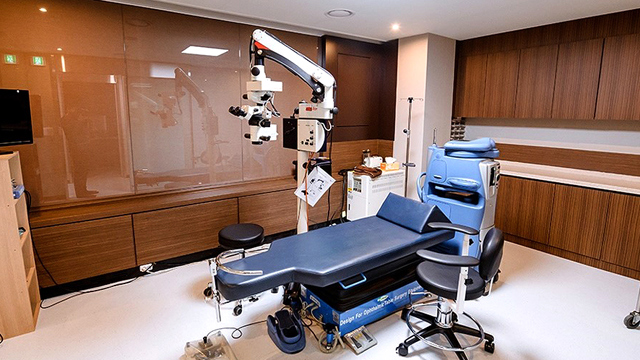
To have new or changing floaters evaluated, contact Jamsil Samsung Eye Clinic in Songpa-gu, Seoul, through its online consultation and contact page or by phone at +82-2-424-7523.
Disclaimer
This page is for general informational purposes and does not constitute individualised medical advice. Diagnosis and treatment recommendations can only be determined through evaluation by a qualified physician.
Other Services
Explore additional ophthalmology treatments offered by our specialists.

EVO AQUA ICL (Implantable Collamer Lens)
EVO AQUA ICL is a vision-correction procedure in which a specialized corrective lens made of biocompatible Collamer material is inserted directly between the natural lens and the iris, without damaging or reshaping the cornea.
Learn More

Presbyopia Surgery (Clareon PanOptix Pro)
Presbyopia surgery with the Clareon PanOptix Pro trifocal lens corrects presbyopia — the age-related loss of near-focusing ability caused by the eye's lens stiffening and losing elasticity, combined with reduced ciliary muscle function — by implanting a
Learn More
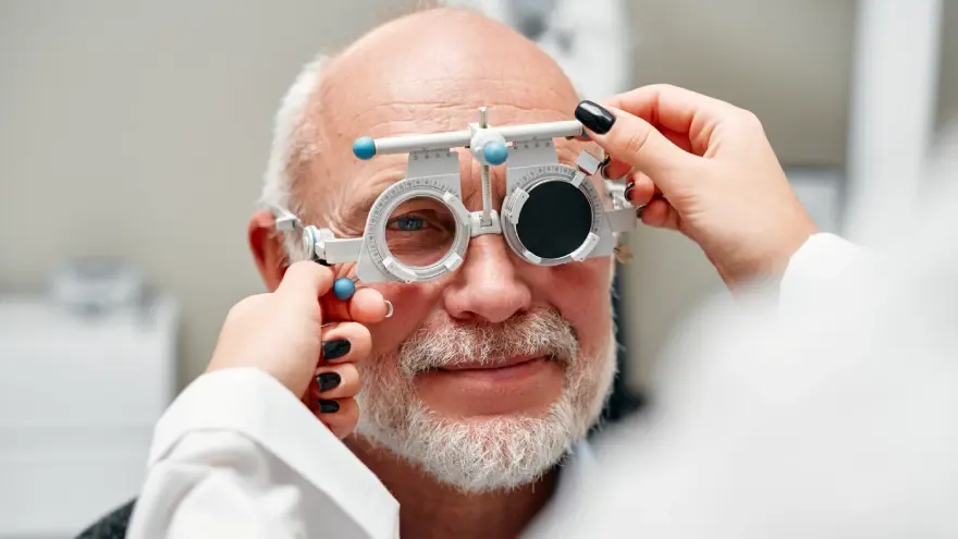
Presbyopia Precision Examination Program
The Presbyopia Precision Examination Program is a specialized diagnostic evaluation designed to thoroughly assess eye health, corneal condition, and refractive index before prescribing presbyopia correction or laser vision procedures.
Learn More