Specialized Ophthalmology in Seoul · Dedicated International Patient Concierge
MOHW Registered · Foreign Patient Care
en-US

Medical Guide
Inside the Pre-Op Exam for LASIK in Seoul: Two Hours
A pre-op exam for LASIK in Seoul runs about two hours. Here is what every instrument measures, why it takes that long, and how the hours after drops feel.
Schedule a ConsultationInstrument by Instrument: The Two Hours That Decide Candidacy
Medically reviewed by Dr. Kim Yoon Kyung, Board-certified ophthalmologist
A pre-op exam for LASIK in Seoul is not a formality you sit through on the way to a procedure. At Jamsil Samsung Eye Clinic it is the point at which eligibility is actually decided, and the record puts it at roughly two hours. Patients who expect twenty minutes and a handshake are often surprised — first by the length, then by how much of it involves machines they have never encountered.
This article walks through what fills those two hours, one instrument at a time: what each device measures, why that measurement matters to a surgical plan, and what it feels like from the chair. Knowing the shape of the morning makes it far less tiring.
Why the decision waits for this room
The National Eye Institute is explicit that suitability for refractive surgery is established through a comprehensive dilated eye exam. Not a questionnaire and not a prescription — an examination.
The reason is that laser correction is a subtractive operation on a specific piece of tissue. Reshaping the cornea, the transparent dome at the front of the eye, means removing a calculated amount of material from a structure whose thickness, curvature and biomechanical behavior differ from person to person. A prescription tells you how much correction is needed. It says nothing about whether this particular cornea can supply it and remain stable.
The order of the tests is not arbitrary either, and it explains the shape of the appointment. Anything that depends on your pupil behaving normally has to happen before the dilating drops go in, because those drops hold the pupil open artificially for hours. Pupil measurement, topography and refraction therefore come first; the interior examination comes last. That is why the appointment cannot simply be shortened by reordering it, and why arriving late costs more than the minutes lost.
Everything in the next two hours exists to answer one of three questions. What correction do your eyes need? Does your cornea have the physical capacity to deliver it? And is there anything present — on the surface, inside the eye, or at the back — that would make the operation unwise regardless?
The opening measurements: acuity and refraction
The examination begins conventionally, with the parts you will recognize from any optometry visit.
Visual acuity is measured first, usually each eye separately, unaided and then with correction. The National Eye Institute lists acuity among the standard components of a comprehensive eye exam, along with visual field testing, eye-muscle function, and how the pupils respond to light. These early tests are quick, and they establish a baseline against which everything later is read.
Eye-muscle function and visual field belong to this early group as well. Muscle testing is simply following a target with your eyes while a clinician watches how the two move together; field testing checks that your peripheral awareness is intact. Neither is altered by refractive surgery, which is precisely why both are recorded now — a baseline captured before an operation is worth far more than one reconstructed after it.
Refraction follows, typically twice. An automated instrument gives a first estimate in seconds — you rest your chin, look at a target, and the machine calculates. Then a clinician refines it manually with a lens bank, asking which of two options looks clearer. The manual step matters because the automated reading is a starting point, not a verdict, and small disagreements between the two are informative in themselves.
MedlinePlus, the National Library of Medicine's consumer service, notes that refractive errors are diagnosed by comprehensive examination, often using dilating drops, and that more than 150 million Americans have one. What varies between those people is not only the strength of the error but its type and how consistently it can be measured. Consistency is part of what is being assessed here.
Corneal topography: a map, not a number
This is the centerpiece, and the test most people have never had.
A curvature reading of the kind used for fitting contact lenses gives a couple of numbers for the central cornea. Corneal topography does something else entirely: it produces a map. The instrument projects a pattern of illuminated rings onto the tear film, photographs the reflection, and computes curvature at thousands of points across the surface. The output is a color-coded chart in which steeper zones and flatter zones are immediately visible.
Two corneas can share an identical prescription and look nothing alike on that map. One may be regular and symmetrical. The other may have a zone of steepening below center, or an asymmetry between the upper and lower halves — features that no number in a spectacle prescription would ever reveal.
That pattern-reading is why the test carries so much weight. The FDA's guidance on when LASIK is not appropriate names keratoconus, a progressive thinning and bulging of the cornea, among the conditions calling for particular caution, and topography is the instrument through which early irregularity of that kind becomes visible. The clinic's record describes mapping both the front and the back surfaces of the cornea, which matters because rear-surface changes can appear before the front surface shows anything.
The test itself is undemanding. You place your chin on a rest, open your eyes wide, and hold still for a few seconds while the image is captured. It is repeated to confirm the capture was clean. Nothing touches your eye and nothing is uncomfortable.
One condition is absolute, and it is set before you arrive: your cornea has to be in its natural shape. Contact lenses mold the surface they sit on, so the clinic requires lenses to have been out for an interval that varies by lens type, with rigid and overnight reshaping lenses needing considerably longer than soft ones. A map produced too early describes the lens's handiwork rather than your eye.
Corneal thickness, and where it is measured
Thickness is the constraint that most often decides how much correction is safely available.
Reshaping removes tissue. What must remain afterward is a bed of cornea substantial enough to hold its form under the eye's internal pressure for the rest of your life. So the arithmetic is straightforward in principle: starting thickness, minus tissue used for the correction you need, minus the layer that must remain untouched.
Thickness is not measured at one spot. Modern imaging generates a thickness map across the whole cornea, and the relationship between the center and the periphery is itself a finding — an unusually thin zone that does not sit centrally can mean something different from uniform thinness.
Two practical consequences follow, and they are worth understanding before anyone explains them to you in a consulting room. A cornea that is thin relative to the correction required may make a surface-based technique more appropriate than a flap-based one, because the two use tissue differently. And thickness limits are a reason a strong prescription can be harder to treat than a mild one in the same eye. The FDA lists thin corneas plainly among the findings that screening looks for.
Pupillometry: measuring the eye in the dark
Pupil measurement is the test whose purpose patients most often miss, and it may be the one that predicts most about how satisfied you are at night.
Your pupil is small in bright light and opens in dim conditions. Laser correction is applied over a treatment zone of finite width. If your pupil in darkness opens wider than that zone, light entering through the untreated periphery is focused differently from light passing through the middle — and that mismatch is one mechanism behind halos around headlights, starbursts from streetlights, and reduced clarity when driving after dark.
This is why the measurement has to be made in the dark, or under controlled dim illumination, rather than in a lit room. A pupil measured under office lighting tells you almost nothing about the pupil you actually drive with. The clinic's record specifies pupil-size measurement under different lighting conditions, and the plural is the point: the useful information is the range between your bright-light and dark-adapted diameter, not a single figure.
The FDA lists large pupils in dim light among the screening findings that weigh against surgery. MedlinePlus separately notes that glare and halos are among the symptoms refractive errors themselves produce, which is why a clinician may ask what you already notice at night before the measurement, and compare the two.
Expect to sit in a darkened room for a few minutes first. Dark adaptation is not instant, and a reading taken too soon understates how wide your pupil goes.
The surface, the tear film, and the lids
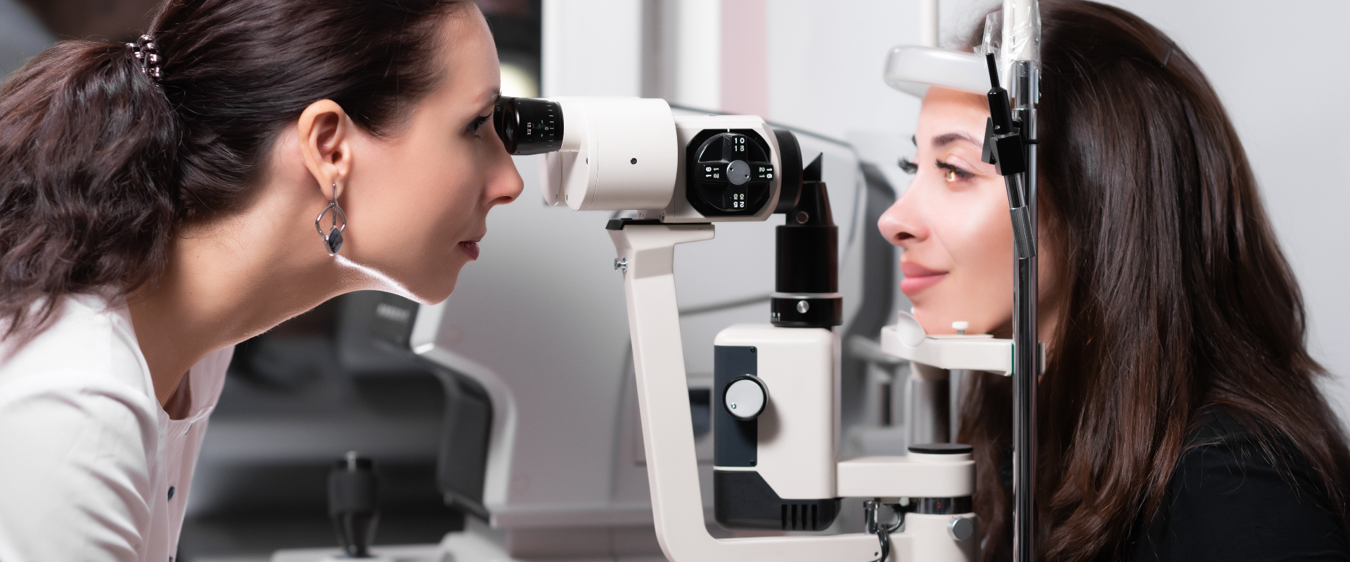
Much of the second half of the examination happens at the slit lamp — a microscope with a narrow, adjustable beam of light that lets a clinician section the eye optically and inspect it layer by layer.
Three things are being assessed here. The clarity and integrity of the cornea itself, including any scarring or irregularity. The tear film, the thin fluid layer coating the surface, and whether it stays intact between blinks or breaks up early. And the eyelid margins, where the oil glands that stabilize that film are located.
Dry eye deserves its own note, because it is both a screening finding and a predictor. The FDA's guidance states that dry eye is among the conditions screening looks for, and that LASIK has a tendency to make it worse. A patient with a marginal tear film before surgery is not in the same position afterward as one with a robust film. This assessment is therefore not a box-ticking exercise; it shapes both the recommendation and what you are told to expect during recovery.
Blepharitis — inflammation of the eyelid margins, also on the FDA's list — frequently produces few symptoms and is found at the slit lamp rather than reported by the patient. It is often manageable, and finding it may mean treating it first rather than abandoning the plan.
Pressure, dilation, and the interior
Eye pressure is measured next. The National Eye Institute includes tonometry in its account of a comprehensive dilated exam, and it is quick: either a brief puff of air, or a probe touching a numbed cornea for a moment. Pressure matters because glaucoma, which the FDA names among conditions requiring extra caution, is associated with it, and because the surgery alters how future pressure readings must be interpreted.
Then come the dilating drops, and the pace of the appointment changes. The drops widen your pupil so the inside of the eye can be examined properly, and they take roughly twenty to thirty minutes to work fully. This is the interval that makes the examination two hours rather than one, and it is unavoidable.
Once dilated, the examination turns to the retina at the back of the eye and to the optic nerve, both of which the clinic's record lists. Neither is reshaped by refractive surgery, but both need to be intact. A retinal thinning or tear found here changes the sequence — some findings are addressed before any refractive procedure is considered — and it is far better found now than after.
The hours after the drops
Plan the rest of the day around this rather than against it.
The National Eye Institute is clear that dilation leaves vision blurry and eyes light-sensitive for a few hours afterward, and advises arranging a ride and bringing sunglasses. Near focus is affected most; text on a phone becomes difficult, while walking around is manageable. Outdoors, ordinary daylight feels harsh.
The clinic states the same in operational terms: because pupils are dilated, you do not drive yourself, and you leave by public transport or with someone accompanying you. Seoul's public transport network reaches the clinic's Songpa-gu premises, so the practical decision is simply made in advance rather than in a bright lobby with dilated pupils. Sunglasses are worth having on you, not in a hotel room across the city.
Do not schedule anything demanding afterward. A meeting, a long walk through an unfamiliar neighborhood, or a museum will all be less pleasant than you imagine. An afternoon with nothing in it is the right plan. You can read more about the practice on its about page.
The conversation at the end
The measurements are only half the value. The examination closes with a discussion, and at this practice the record describes a one-to-one arrangement in which the operating surgeon handles pre-operative and post-operative care personally — so the person interpreting these results is the person who would perform the procedure.
Four things are worth getting from that conversation, and it is reasonable to write them down. Which findings, specifically, drive the recommendation you are given. Whether any measurement was borderline or needed repeating. What your night vision profile suggests, in light of your pupil measurement and what you already notice. And what the alternative would be if the first recommendation does not suit you.
You may also be told that a decision cannot be reached today — because a reading was inconsistent, because a finding needs treating first, or because something has to be remeasured. The FDA's own account of the process notes that measurements are sometimes repeated at a later visit. That is the examination doing its job. Accounts from other patients, including those on the clinic's reviews page, describe the exam day as substantial; treat that as the expectation rather than the exception.
The Bottom Line
- The examination runs about two hours because it is a sequence of distinct tests, and because dilating drops need twenty to thirty minutes to take effect before the interior of the eye can be assessed.
- Corneal topography maps curvature at thousands of points rather than reporting a single figure, which is how irregularity invisible in a prescription becomes apparent. Corneal thickness then sets how much correction is physically available.
- Pupil size is measured under varying illumination because a pupil that opens wider than the treated zone in darkness is linked to halos and glare at night.
- Dilation leaves vision blurry and light-sensitive for a few hours. The National Eye Institute advises arranging a ride and bringing sunglasses, and the clinic requires that you not drive yourself.
- Final eligibility is settled here, not before. A deferred or negative answer means the examination worked.
Frequently Asked Questions
Does the whole examination really take two hours?
The clinic's record describes a comprehensive examination of roughly that length. Active testing accounts for part of it; a meaningful share is waiting for dilating drops to take effect, plus the closing discussion of results. Allow the full morning or afternoon rather than trying to fit it between other commitments.
Which of the tests are uncomfortable?
Very few. Topography, thickness imaging and pupil measurement involve no contact with the eye at all. Tonometry is either a brief puff of air or a momentary touch on an anesthetized cornea. Dilating drops sting for a second or two. The most trying part for most people is the light sensitivity afterward, not any individual test.
Why measure my pupils in a dark room?
Because the pupil you drive with at night is the relevant one. In office lighting the pupil is constricted, and a measurement taken there understates its dark-adapted size. If it opens wider in darkness than the treated area of the cornea, light passing through the untreated edge is focused differently, which is associated with halos and glare after dark.
What happens if a finding rules out the procedure?
It depends on the finding. Some are temporary and treatable — inflamed eyelid margins, for instance, may be managed and the assessment revisited. Some call for a different technique rather than none. Others, such as evidence of corneal thinning disease, are lasting reasons not to proceed. The FDA likewise names a recent shift in prescription, inside the past year, as grounds for postponement rather than permanent exclusion.
Can I go straight from the examination to surgery the same day?
The clinic documents same-day evaluation-to-procedure schedules for qualifying candidates, so it is possible in principle. Whether it applies to you is exactly what the examination determines, and it is not something to assume in advance. Ask about it directly rather than building a travel plan around it.
Will I be able to see well enough to get back on my own?
You will be able to walk and navigate, but near vision will be poor and bright light will be uncomfortable for a few hours. Self-driving is ruled out because of dilation. Public transport or a companion is the expected route, and sunglasses make the trip considerably easier.
Why is my old prescription not enough to decide this?
Because a prescription describes the correction your eye needs, not the tissue available to deliver it. Two people with identical prescriptions can have very different corneal thickness, curvature patterns and pupil behavior — and those differences determine what is safe. The National Eye Institute places the suitability decision on a comprehensive dilated exam for that reason.
This article is for general informational purposes and does not constitute individualized medical advice. Only a qualified ophthalmologist who has examined your eyes can determine whether any refractive procedure is appropriate for you. It is not a self-diagnosis tool.
More Articles
Continue reading our clinical ophthalmology guides.
LASIK Cost Korea vs US: How Each System Sets Prices
LASIK cost in Korea vs the US differs because each system treats vision correction as elective. How pricing is structured, and how to request a real quote.
Learn More
Dr. Kim Byoung Jin: Ophthalmologist in Seoul, Korea
Dr. Kim Byoung Jin is a board-certified ophthalmologist and representative director at Jamsil Samsung Eye Clinic, Seoul. His training and focus, explained.
Learn More

Book an Online Consultation: Korea Eye Clinic Guide
Book an online consultation with a Korea eye clinic: the channels this Seoul practice documents, the time-zone arithmetic, and what to assemble up front.
Learn More